High School Workshop
Have you ever felt that plants are less exciting than animals? While animals show rapid, visible movement, plants can seem still; but at the cellular level, they are surprisingly active. With a microscope, even the most stationary plants reveal dynamic movement at the microscale.
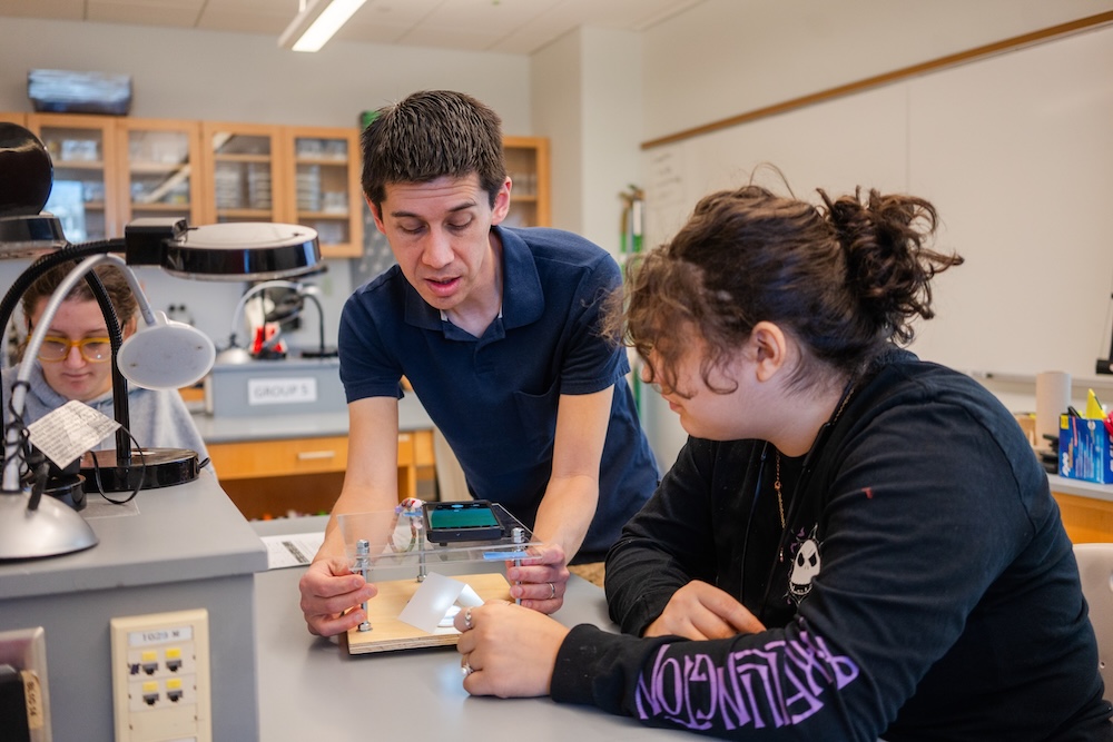
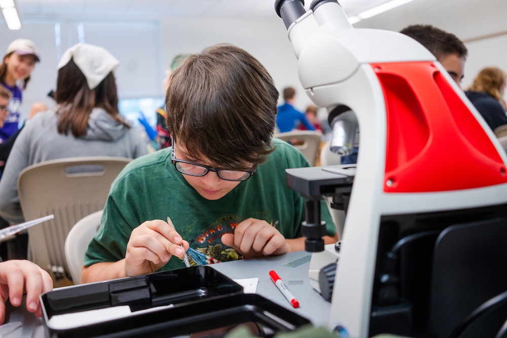
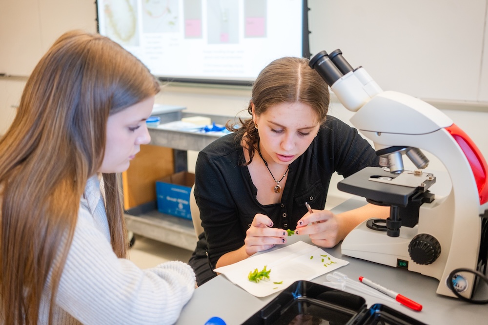
In this session, students explore light-induced chloroplast movement in aquatic plants. They develop hypotheses about underlying mechanisms and design experiments to test their ideas. Students use both compound and simple microscopes to investigate the microscopic world. By the end, they gain a deeper appreciation for plant activity at the cellular level and for the process of scientific inquiry.
Faculty Developer: Dr. Chris Kubow, Biology


What students do:
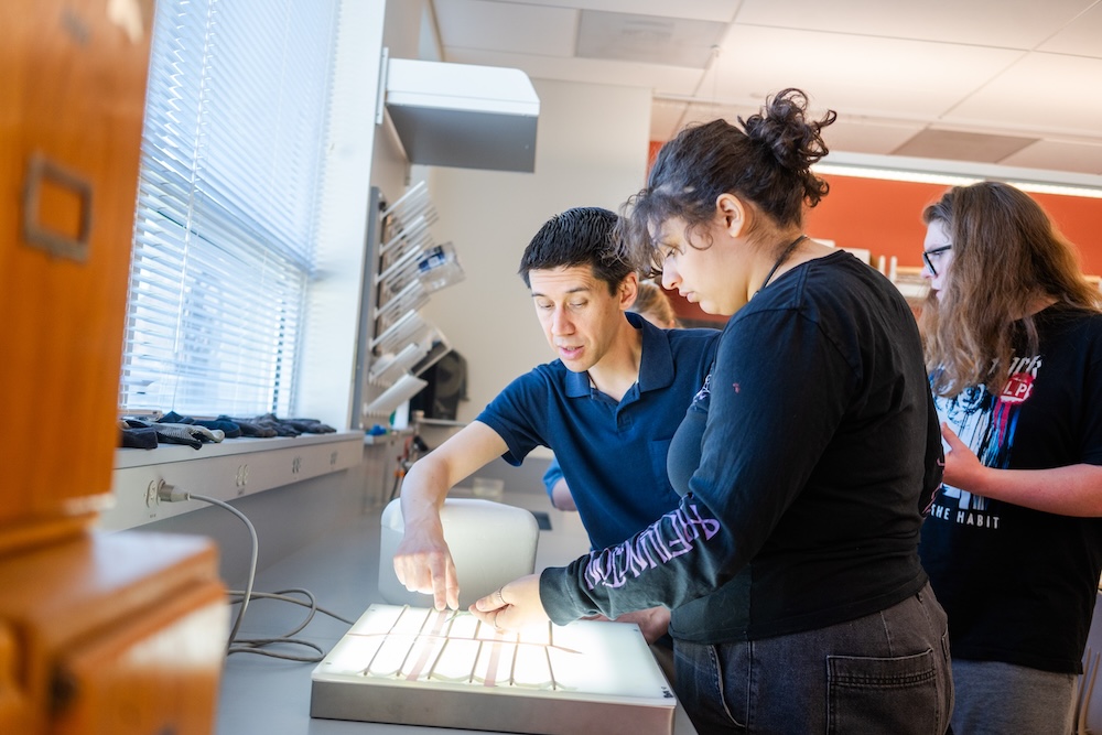
After a brief introduction to scientific discovery, students work in groups to develop a hypothesis, design an experiment, and predict outcomes related to chloroplast movement. They then conduct their experiment by preparing samples, exploring different microscopes, and analyzing their results. Finally, as a class, they draw and share their conclusions.
What students learn:
- Understanding the process of scientific discovery
- How to develop a hypothesis and design an experiment to test it
- Mechanisms behind chloroplast movement
- How to prepare microscope samples
- Use of compound and minimalist microscopes
- Drawing conclusions based on experimental results
- The importance of cell movement, including its role in cancer
- The value of simple, low-cost microscopes for science and healthcare in underserved regions
Background Knowledge
- Parts of a eukaryotic cell
- Scientific inquiry cycle
- Basics of photosynthesis
Information for Teachers:
- A short article describing two types of chloroplast movement in Elodea.
- Bob Goldstein (UNC Chapel Hill) website featuring microscopes like those used in our workshops.
Connection to research:
Dr. Kubow studies cell movement in tissue. While his work focuses on mammalian cells, many of the same molecules and processes also drive movement in plant cells. He directs JMU’s Light Microscopy Facility, supporting research with advanced imaging systems.